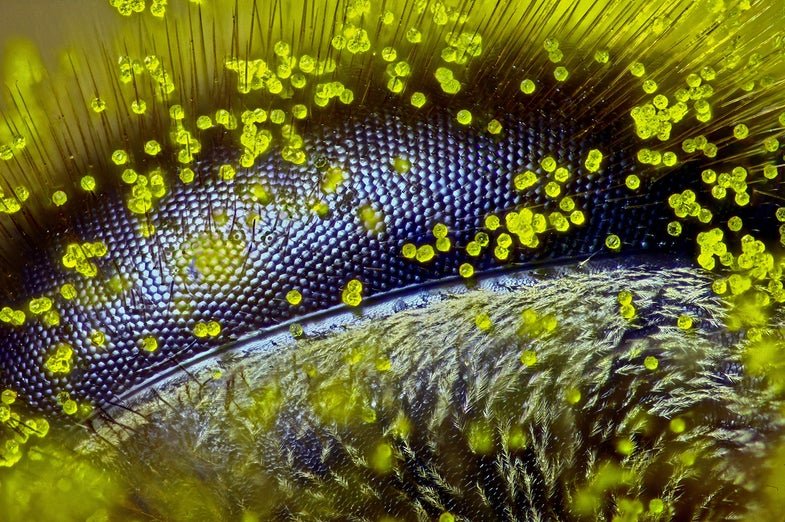
There are plenty of large, incredible things in this world, from canyons to oceans and huge fossils left behind by dinosaurs. But most of the things that make up our wide world are small–tiny even. Often, these things escape notice because they are simply to small for the human eye to see. Which is where Nikon’s annual Small World competition comes in. Every year a panel of judges selects the most stunning images of very small things. Often, the people who capture these images are scientists, who come across these stunning images in the course of their daily work. Nikon received over 2,000 submissions from 83 countries for this year’s competition.
Second Place: Mouse Colon
Gut microbes are of increasing interest to scientists, who are finding that the microbes who live inside us play an outsized role in our health. This is an image of a mouse colon colonized with human gut microbes.
Third Place: Bladderwort
Open wide! This is the intake, or entrance to the interior of a bladderwort, a carnivorous freshwater plant. The plant sucks prey into its trap, where it becomes a tasty meal. But don’t worry, it isn’t Audrey II. This little guy is less than 2 millimeters long.
Fourth Place: Mammary Gland
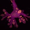
The pink blob in this image is a lab-grown human mammary gland organoid. Researchers have grown breast tissue in the lab to better understand how cancer forms, and also how mammary glands originated.
Fifth Place: Brain Tumor
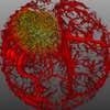
Researchers imaged this picture of a growing glioblastoma (brain tumor) in a mouse. The red ‘netting’ surrounding the tumor is part of the mouse’s vascular (blood supply) system.
Sixth Place: Moss
The spore capsule on a tiny bit of moss.
Seventh Place: Starfish
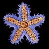
This image of a starfish has been enlarged 10 times under a microscope.
Eighth Place: Mouse Ear
The ear of a mouse is an incredibly soft and delicate thing. In this magnified image you can see the nerve and blood vessels going through the ear.
Ninth Place: Flower Buds
These aren’t buds you’d want to put in a vase, but they are the very beginning of life for a flowering plant called Arabidopsis, which is related to cabbage and mustard. This image was taken at 40x magnification.
Tenth Place: Clam Shrimp
A live clam shrimp photographs beautifully when prepared for its close-up.


