Every year since 1974, Nikon has herded a gaggle of nerds into a room for an impossible task: Pore over thousands of microscope images and pick the very best ones.
I was lucky enough to join the judge’s circle this summer for Nikon Small World 2014—the 40th year of the microscopy competition. Our small group had to peruse more than 1,200 entries from 79 countries.
It wasn’t easy. Many of the images had just the right mix of contrast and color to make them nearly leap off the screen. But our judging criteria went beyond visual allure; a gorgeous-looking image can be surprisingly commonplace while a mysterious-looking shot is borderline revelatory. So, we selected the finest shots based not only on appearance but also creativity, informational content, and technical execution.
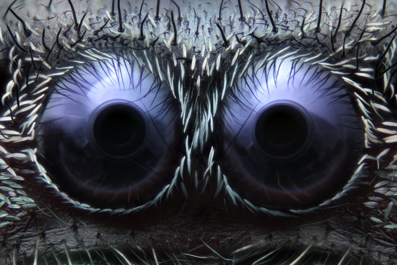
The 20 images you’re about to see (including Mr. Jeepers Creepers jumping spider eyes, above) really are the best. There’s everything from psychedelic-looking algae and frighteningly detailed caterpillar feet to hyperrealistic rotifers and rainbow-colored worm babies.
One more thing before you dive in: If you find yourself curious about the techniques used to make these award-winning images, I’d recommend a visit to Molecular Expressions and MicroscopyU. These sites aren’t exactly light reading, but they do host countless interactive visuals—including this animation, which demystifies the complex, Nobel Prize-winning superresolution microscopy method—that go a long way in helping you understand how the techniques work.
Click an image below to start blowing your mind.
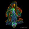
Crawling Bone Cancer (20th Place)
This is the front part of a cell of bone cancer, or osteosarcoma, that’s crawling using protein-based fibers called actin. The rainbow of color is added later to sort out the cell’s complex structure. View the high-resolution version Technique: Structured illumination microscopy Magnification: 8000X
Acorn Worm Larva (19th Place)
Shown here is an acorn worm larva, also known as Balanoglossus misakiensis, from above. The false color highlights cell borders, muscles, and even eye spots. View the high-resolution version Technique: Confocal microscopy Magnification: 10X
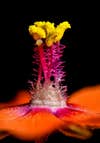
Scarlet Pimpernel (18th Place)
Famous as the symbol for Emma Orczy’s turn-of-the-century novel, a scarlet pimpernel (or Anagallis arvensis) has a bundle of stamen that rises like a tower in this macro photograph. View the high-resolution version Technique: Macroscopy Magnification: 80X
Transgenic Kidneys (16th Place)
This extreme close-up shows three cultures of transgenic kidneys grown together. Red and blue-green colors highlight collisions of branching collecting duct systems. View the high-resolution version Technique: Confocal microscopy Magnification: 20XNils
Agate Mineral (12th Place)
An unpolished slab of Montana Dryhead agate reveals rich yellow and red minerals. View the high-resolution version Technique: Axial lighting microscopy Magnification: 50X
Caterpillar Foot (4th Place)
This terrifying orifice isn’t the mouth of a beast, but rather the stubby little leg and foot of a caterpillar. The hooks reveal how the insect larvae manage to stick to almost any surface. (We dare you not to think of how scary a caterpillar’s foot is the next time you pick one up.) View the high-resolution version Technique: Confocal and autofluorescence microscopy Magnification: 20X
Jumping Spider Eyes (3rd Place)
This soul-chilling pair of eyes belongs to a common jumping spider. “I find that looking directly into a spider’s front eyes is very powerful, as it’s a perspective many of us aren’t used to,” says 19-year-old Noah Fram-Schwartz. Schwartz created this photo by splicing together 80 different microscope images, which have a very thin plane of focus, into one ultra-crisp image. View the high-resolution version Technique: Reflected light microscopy Magnification: 20X
Calcite Crystal (2nd Place)
Crystals love to grab and scatter light into a blinding glow under a microscope. But with a little polarized light, their angles and surface detail emerge in sharp relief. This is a piece of calcite, which is one of the most common minerals on Earth, yet occurs in a seemingly unlimited variety of shapes and colors. One thing all calcite crystals have in common, however, is perfect rhombohedral cleavage—a slanted-cube structure shown clearly in this image. View the high-resolution version Technique: Cross-polarized light microscopy Magnification: 10X
Swimming Rotifer (1st Place)
Although Rogelio Moreno of Panama only began his adventures in microscopy about five years ago, he’s become a regular contender in the Nikon Small World competition. This year, however, he finally took first prize. Shown here is Moreno’s rare shot of a high-speed rotifer caught open-mouthed and facing the camera, revealing its red, heart-shaped corona. Moreno babysat his microscope for hours on end for the right moment. When it came, he snapped a photo using a special beam-splitting strobe light—and froze this busy rotifer in place in sharp, almost 3-D–like relief. “Rotifers are very difficult to photograph,” says Moreno. “I’ve always wanted to take this photo, but you have to wait and wait and wait.” View the high-resolution version Technique: Differential interference contrast microscopy Magnification: 40X
