Gallery: The Best MicroPhotography of 2011
Nikon announced the winners of its Small World microphotography contest and once again, the results are mesmerizing
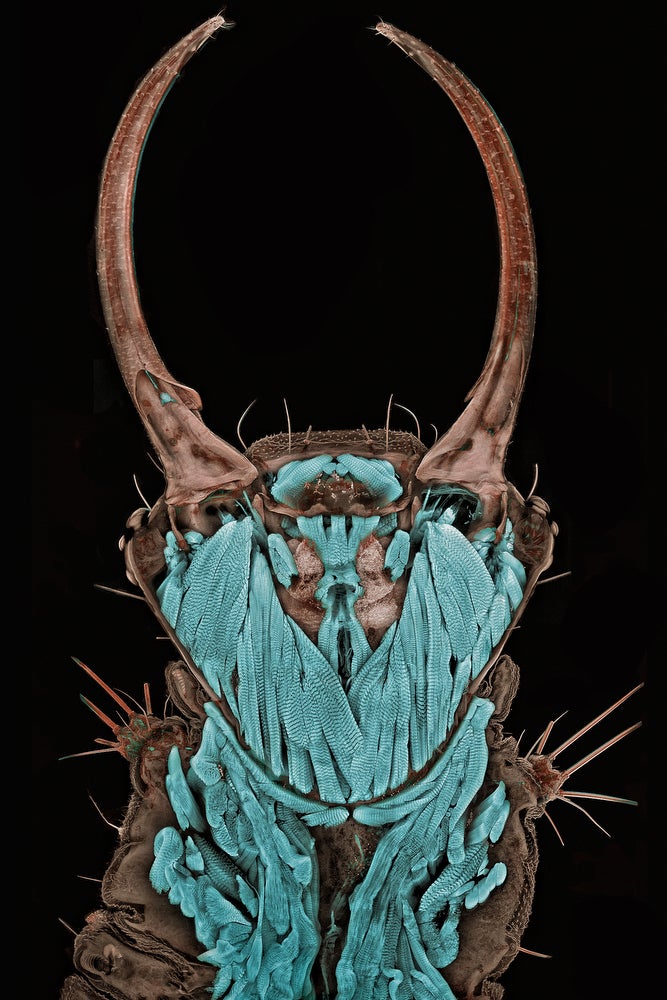
Photo: Dr. Donna Stolz
University of Pittsburgh
Pittsburgh, Pennsylvania, USA
Blade of Grass (200X)
Confocal stack reconstruction, Autofluorescence
Photo: Frank Fox
Fachhochschule Trier
Trier, Rheinland-Pfalz, Germany
Melosira moniliformis, living specimen (320X)
Differential Interference Contrast
Photo: Dr. Robin Young
The University of British Columbia
Vancouver, British Columbia, Canada
Intrinsic fluorescence in Lepidozia reptans (liverwort) (20X)
Live mount, Confocal microscopy
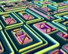
Photo: Alfred Pasieka
Germany
Microchip surface, 3D reconstruction (500X)
Incident light, Normarski Interference Contrast
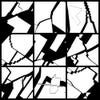
Photo: Dennis Callahan
California Institute of Technology
Pasadena, California, USA
Cracked gallium arsenide solar cell films (50X)
Brightfield
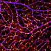
Photo: Gabriel Luna
UC Santa Barbara, Neuroscience Research Institute
Santa Barbara, California, USA
Retinal flatmount of mouse nerve fiber layer (40X)
Laser Confocal Scanning

Photo: Dr. Bernardo Cesare
Department of Geosciences
Padova, Italy
Graphite-bearing granulite from Kerala (India) (2.5X)
Polarized light
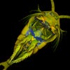
Photo: Dr. Jan Michels
Christian-Albrechts-Universität zu Kiel
Kiel, Germany
Temora longicornis (marine copepod), ventral view (10X)
Confocal, Autofluorescence and Congo Red Fluorescence
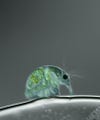
Photo: Joan Röhl
Institute for Biochemistry and Biology
Potsdam, Germany
Daphnia magna (freshwater water flea) (100X)
Differential Interference Contrast
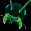
Photo: Dr. Jan Michels
Christian-Albrechts-Universität zu Kiel
Kiel, Germany
Ant head, frontal view (10X)
Confocal, autofluorescence
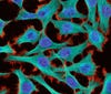
Photo: Thomas Deerinck
National Center for Microscopy and Imaging Research
La Jolla, California, USA
HeLa (cancer) cells (300X)
2-Photon fluorescence
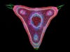
Photo: Dr. Stephen S. Nagy
Montana Diatoms
Helena, Montana, USA
Curare vine in cross-section, Chondrodendron tomentosum (45X)
Brightfield, digitally inverted
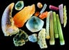
Photo: Yanping Wang
Beijing Planetarium
Beijing, China
Sand (4X)
Reflected light
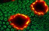
Photo: James H. Nicholson
Coral Culture and Collaborative Research Facility, NOAA/NOS/NCCOS/CCEHBR & HML
Charleston, South Carolina, USA
Porites lobata (lobe coral), live specimen displaying tissue pigmentation response with red fluorescence (12X)
Epifluorescence with triple band (U/B/G) excitation
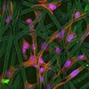
Photo: Dr. Christopher Guérin
VIB (Flanders Institute of Biotechnology)
Ghent, Belgium
Cultured cells growing on a bio-polymer scaffold (63X)
Confocal
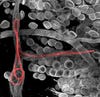
Photo: Dr. Witold Kilarski
EPFL-Laboratory of Lymphatic and Cancer Bioengineering
Lausanne, Switzerland
Litomosoides sigmodontis (filaria worms) inside lymphatic vessels of the mouse ear (150X)
Fluorescent confocal microscopy
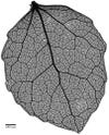
Photo: Benjamin Blonder, David Elliott
University of Arizona
Tucson, Arizona, USA
Venation network of young Populus tremuloides (quaking aspen) leaf (4X)
Brightfield image of safranin-stained tissue
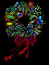
Photo: Dr. Donna Stolz
The University of Pittsburgh
Pittsburgh, Pennsylvania, USA
Mammalian cell collage stained for various proteins and organelles, assembled into a wreath (200-2000X)
Single slice confocal cell mosaic
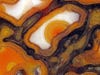
Photo: Douglas Moore
University of Wisconsin – Stevens Point
Stevens Point, Wisconsin, USA
Agatized dinosaur bone cells, unpolished, ca. 150 million years old (42X)
Stereomicroscopy, fiber optics